
Blog
Fingernails, a keratinized protein like hair, are emerging as a popular specimen type for alcohol and other drug testing. Although fingernail specimens have been used for toxicological analysis and pharmacokinetic studies for decades, many people have less experience interpreting the results than hair and urine, and there is confusion about whether to use nail clipping or scraping. 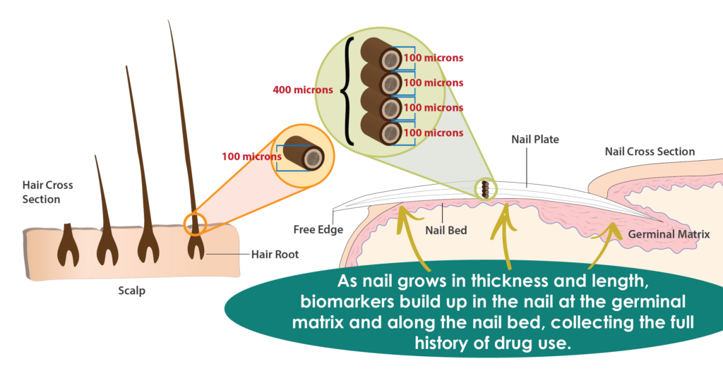
How Far Back Does Fingernail Testing Go?
One of the most frequent questions we receive revolves around the detection window for fingernail testing. To understand the detection window, we need to first discuss the anatomy of the fingernail and the incorporation of compounds in the nail first.
Anatomy of the Nail
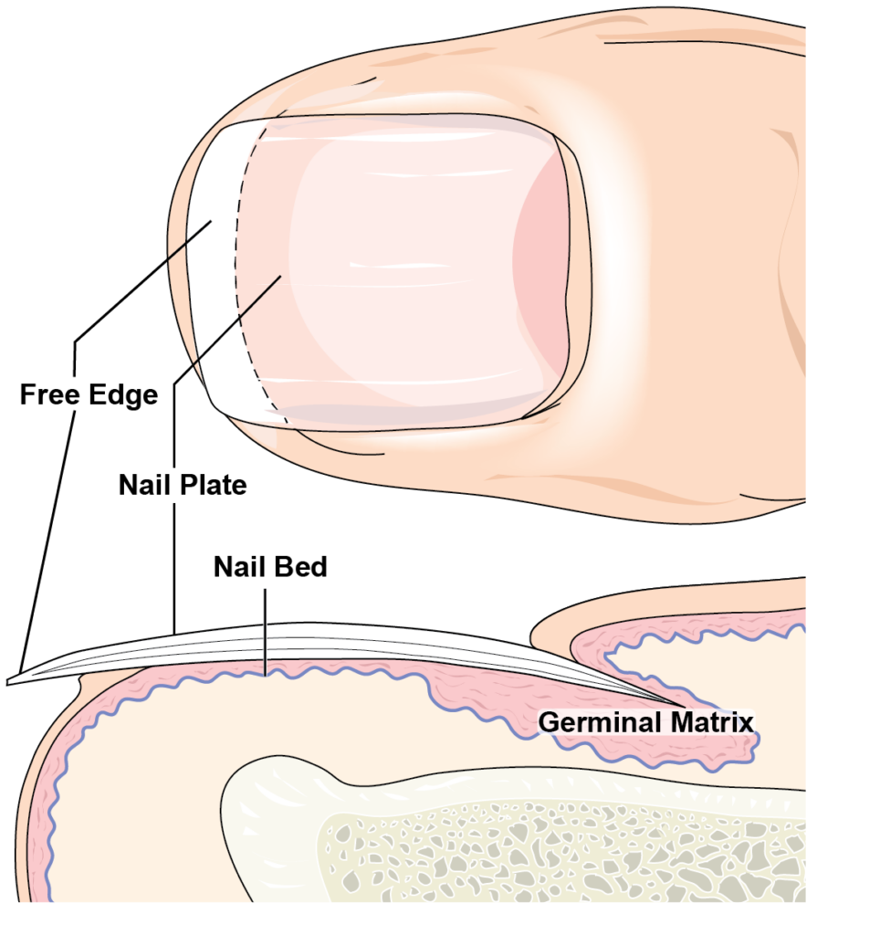
Nail is a keratinized protein very similar to hair. It is porous, and compounds become trapped and bind within the structure. For this discussion, we need to know four anatomical features of the nail: the germinal matrix, the nail plate, the nail bed, and the free edge (Figure 1). The nail originates at the germinal matrix and grows outward toward the fingertip. The hardened material forming the nail plate grows across the nail bed, which is rich in capillary blood flow (this causes the pinkish color to your fingernails). As the nail grows, material is added from underneath, lengthens, and thickens as it grows outward toward the fingertip. Once the nail plate erupts from the nail bed and extends past the end of the fingertip, it is called the free edge. The free edge is the piece that you clip cut off when clipping your nails. The entire process takes up to 6 months, depending on the individual’s health.
The 4 Routes of Biomarker Incorporation
Compounds are incorporated into the nail by four main routes. The first route, just like hair, is environmental exposure. If someone is handling a drug or near around someone smoking a drug, the drug gets on the nail, works its way into the pores, and binds to the keratinized protein. The second route is the sweat and oil of the skin surrounding the nail, which deposits drugs and drug metabolites into the nail. The third route of incorporation is the blood flow in the germinal matrix, which deposits drug and drug metabolites into the forming nail. Lastly, drug and drug metabolites are deposited to the underside of the nail plate by the blood flow in the nail bed. These four very different routes of incorporation are superimposed on top of each other, rendering a very complex drug history.
Detection Window
When alcohol or other drugs are ingested, biomarkers can be found in nails as early as 1-2 weeks later. The time period of detection depends on the substance used, the amount used, and personal metabolism. Given the anatomy of nail growth and the incorporation routes, drug and alcohol biomarkers may be detectable in fingernails for up to approximately 3-6 months. Like any drug test, time, dose, or frequency of use cannot be inferred, and a negative result is not proof of abstinence.
Clipping, No Nail Scraping!
Since the clipping contains the entire growth and history of the nail, nail scraping is unnecessary. Fingernail specimens are clipped and collected by the donor under the observation of a trained collection staff member. A clipping of 2-3 mm long (about the width of a quarter) from all ten fingernails will, in most cases, give an acceptable amount for screening and confirmation.
Reference A. Palmeri et al. (2000). Drugs in nail: Physiology, pharmacokinetics, and forensic toxicology. Clinical Pharmacokinetics. 38(2), 95-110.
- Drug Classes and Neurotransmitters: Amphetamine, Cocaine, and Hallucinogens
- Environmental Exposure Testing for Delta-8 THC, Delta-9 THC, Delta-10 THC, and CBD
- Bromazolam and Synthetic Benzodiazepines
- Winter Weather Delay Update
- Tianeptine
- Revolutionizing DUI Interventions: Wisconsin’s Breakthrough in Biomarker Testing for Impaired Drivers
- 3 FAQs You Should Know About Newborn Drug Testing
- The Brain Chemistry Behind Tolerance and Withdrawal
- March 2024 (1)
- February 2024 (1)
- January 2024 (3)
- December 2023 (1)
- November 2023 (1)
- August 2023 (1)